
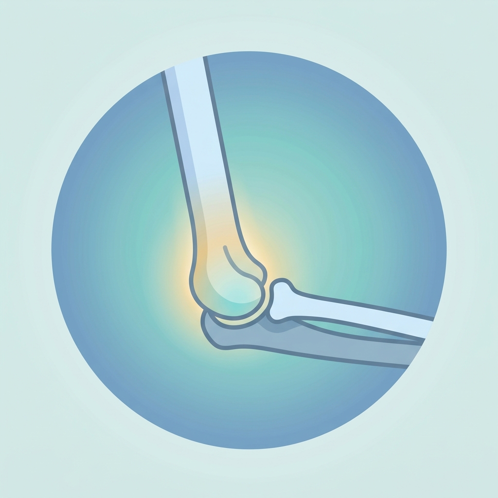
Supracondylar Fracture
Get Clear, Concise Answers to Your Top Questions About Supracondylar Fracture
Q1. What is a supracondylar fracture?
A supracondylar fracture is a break in the humerus (the upper arm bone) just above the elbow joint. This area of the bone is relatively thin, making it a common site for fractures when significant force is applied. It is one of the most frequent elbow fractures diagnosed, particularly in young children.
Q2. Who is most likely to get a supracondylar fracture?
These fractures are most common in children between the ages of 5 and 10. A child\'s bones are still developing, and the thin area of bone just above the elbow is relatively weak compared to the surrounding ligaments and joints. While less common, adults can also experience supracondylar fractures, typically as a result of high-energy, high-impact trauma.
Q3. What causes a supracondylar fracture?
The most common cause is a fall onto an outstretched hand, often from playground equipment such as monkey bars, swings, or trampolines. It can also occur from bicycle falls or during contact sports. The impact forces the elbow to hyperextend, placing immense stress on the humerus bone just above the joint and causing it to fracture.
Q4. What are the common symptoms of a supracondylar fracture?
Symptoms of a supracondylar fracture typically appear immediately after the injury and include:
- Severe and constant pain in the elbow and lower part of the upper arm.
- Rapid and significant swelling, along with bruising around the joint.
- Inability to bend, straighten, or rotate the elbow.
- A visible deformity, making the arm look crooked or bent out of shape.
- Numbness, tingling, or coldness in the hand or fingers, which indicates pressure on nerves or blood vessels.
Q5. How is a supracondylar fracture diagnosed?
A doctor will perform a thorough physical examination to assess the elbow for tenderness, swelling, and deformity. They will also check the pulses in the wrist and assess nerve function in the hand to ensure there is no nerve or blood vessel compression. X-rays are then taken from multiple angles to confirm the fracture, show how much the bones have shifted, and check for any joint misalignment.
Q6. Why is a supracondylar fracture considered a medical emergency?
This fracture is a medical emergency because the sharp edges of the broken bones can compress or injure the nerves and major blood vessels (specifically the brachial artery) that run right past the elbow to the hand. If blood flow is blocked or nerve function is compromised, immediate medical intervention is required to prevent permanent damage to the forearm and hand.
Q7. What are the non-surgical treatment options?
If the fracture is non-displaced (the bone is broken but the pieces are still aligned in their correct position), surgery is generally not needed. The arm is placed in a splint or cast for 3 to 4 weeks to keep the elbow completely still while the bone heals. Regular follow-up appointments and X-rays are scheduled to confirm that the bones remain in proper alignment during recovery.
Q8. When is surgery necessary for a supracondylar fracture?
Surgery is necessary if the bones are displaced (moved out of their normal position), if the fracture is open (bone has broken through the skin), or if there is any evidence of compromise to blood vessels or nerves. Realigning the bones precisely is critical to ensure proper healing, restore normal elbow movement, and prevent long-term growth complications.
Q9. What does supracondylar fracture surgery involve?
The standard surgical treatment is a procedure called closed reduction and percutaneous pinning. While the patient is asleep under general anesthesia, the surgeon carefully manipulates the bones back into their proper alignment without making a large incision. The surgeon then inserts small, smooth metal pins through the skin and into the bone to hold them securely. A cast or splint is then applied over the arm, leaving the pins in place.
Q10. What are the risks of supracondylar fracture surgery?
While this surgery is highly common and successful, potential risks include:
- Infection at the pin sites where the metal pins enter the skin.
- Temporary or permanent nerve injury, which can cause weakness or numbness in the hand.
- Stiffness or a temporary reduction in the elbow\'s range of motion.
- Malunion, where the bone heals in a slightly crooked position, potentially leading to a cosmetic deformity.
Q11. How long does the healing and recovery process take?
The bone typically heals enough for the cast and pins to be removed after 3 to 4 weeks. The pins are removed in the clinic without needing another surgery or anesthesia. Most patients, especially children, regain most of their movement within 6 to 8 weeks, with complete recovery of strength and range of motion taking up to 6 months.
Q12. What should I expect during recovery (cast care, pain management)?
During recovery, it is important to follow these guidelines:
- Keep the cast completely dry. Cover it with a plastic bag or waterproof sleeve during baths.
- Elevate the arm on pillows above the level of the heart, especially for the first 48 to 72 hours, to reduce swelling.
- Use prescribed or over-the-counter pain medications as directed by your doctor.
- Encourage your child to wiggle their fingers frequently to help reduce swelling and monitor circulation.
Q13. Will physical therapy or exercises be needed?
Children rarely require formal physical therapy after cast and pin removal. They naturally regain their elbow motion and strength simply by playing and using their arm for everyday activities. For adults or in complex cases where stiffness persists, structured physical therapy exercises may be recommended to help restore full range of motion.
Q14. What are the potential long-term complications or effects?
Most patients recover completely without any long-term issues. Rare complications can include:
- Gunstock deformity (cubitus varus): A cosmetic change where the elbow angles slightly inward, which rarely affects arm function.
- Growth plate injury: Very rarely, the fracture can affect the growth plate, leading to asymmetric growth of the arm.
- Chronic stiffness: A permanent loss of minor bending or straightening capability, which is more common in adults than children.
Q15. When should I contact my doctor immediately during recovery?
You should contact your doctor or seek immediate emergency care if you notice any of the following symptoms:
- Fingers that become cold, pale, blue, or purple.
- Severe pain that is not relieved by pain medication or elevation.
- New or worsening numbness, tingling, or inability to move the fingers.
- Pus, a foul odor, or increasing redness coming from the cast or around the pin sites.
- A cast that becomes loose, soft, cracked, or wet.
We encourage you to write down any specific questions you have and discuss them thoroughly with your surgeon, doctor, or physical therapist. They are your best resource for personalized advice and care.
Disclaimer: This brochure provides general information and is not a substitute for professional medical advice. Always consult your doctor or qualified healthcare provider with any questions you may have regarding your specific medical condition and treatment plan.

Related articles
Browse all articlesTrusted External Resources
Curated educational materials to help you make informed decisions about your orthopaedic bone & joint conditions.

Orthopaedia
Orthogate Patient Education